3D bioprinting can be used to produce organs and tissues. However, the bio-inks do not adequately replicate the physiological structure and composition of tissues. Fraunhofer researchers have developed bio-printing inks made from the most common tissue proteins to address this issue.
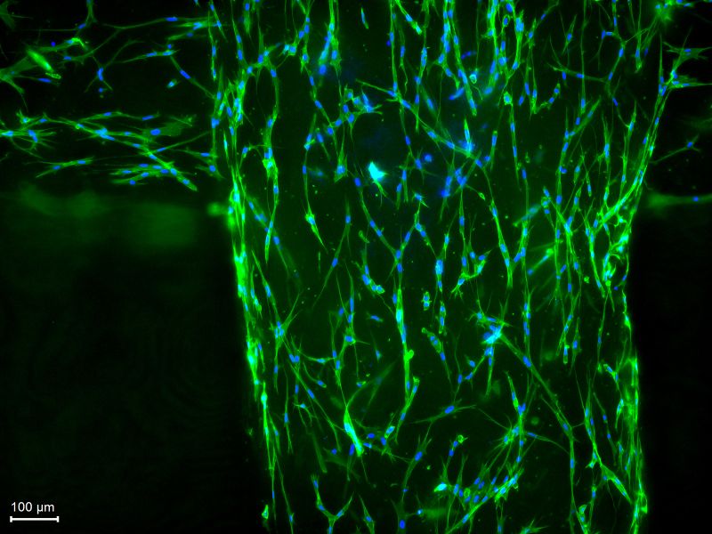
In the PhysioINK project, Fraunhofer researchers are developing bio-printing inks from the most common tissue proteins. Under the microscope, the living tissue cells (fibroblasts) are visible in the printed lattice structure: blue represents the cell nucleus, and green represents the cytoskeleton.
(Source: Tobias Weigel/Fraunhofer ISC)
Every year in Germany alone, 8,000 people are on a waiting list for an organ donation, and about 10 percent of them wait in vain. 3D bioprinting is seen as a potential solution to the global shortage of donor organs. It is intended to enable the automated production of implantable organs or parts of organs, individually tailored to the patient, “at the push of a button.” The technology could also allow for the printing of pathological—that is, diseased—tissue models for disease research as well as for the testing and development of active ingredients. However, one challenge of 3D bioprinting that remains unresolved is the choice of printing inks: some have good printing properties but cannot reproduce the natural tissue structure, while others can reproduce the tissue structure but lack sufficient printability. The Fraunhofer Institutes for Silicate Research ISC, for Applied Polymer Research IAP, for Microstructure of Materials and Systems IMWS, and for Cell Therapy and Immunology, Division of Bioanalytics and Bioprocesses IZI-BB, aim to overcome this hurdle. In the PhysioINK project, they aim to develop sustainable, process-optimized bio-printing inks that enable the precise replication of physiological human organ structures and can be used effectively in various 3D printing processes. The plan is to implement two applications: standardized intestinal tumor models for drug development and physiological heart tissue as a step toward functional implants for personalized, regenerative medicine.
Highly Concentrated Printing Inks Based on Collagen and Elastin
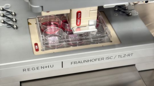
3D Printing of Freestanding Lattice Structures
(Source: K. Selsam/Fraunhofer ISC)
Human soft tissue contains fibrous, structure-forming proteins: collagen and elastin. Depending on the type of tissue, they account for 60 to 100 percent of its composition. “Until now, completely synthetic or heavily chemically modified materials have been used for printing inks. We, on the other hand, use physiological material—that is, the same material that makes up organs,” says Dr. Tobias Weigel, project coordinator and researcher at Fraunhofer ISC in Würzburg (Germany). As a first step, the project team is developing highly concentrated printing inks based on proteins—specifically type I collagen, type IV collagen, and elastin—which are characterized by a neutral pH, physiological properties, and high compatibility with human cells. To achieve this, the consortium combines cellulose sulfates that are molecularly tailored to the specific collagens or structural proteins, which initially stabilize the proteins in their dissolved and printable state. A temperature trigger then reverses this stabilization, allowing the structural proteins to self-assemble into their physiological fibrous or network-like structure.
Encapsulate Positively Charged Collagen Molecules with Negatively Charged Cellulose Sulfates
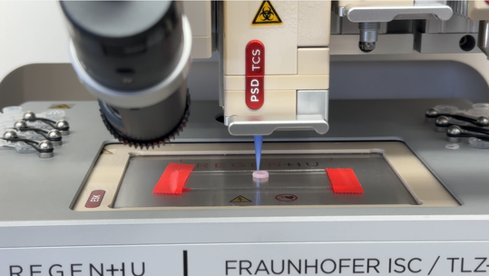
3D Printing of Freestanding Ring Structures
(Source: K. Selsam/Fraunhofer ISC)
As part of the PhysioINK project, researchers are encapsulating positively charged collagen and elastin molecules with corresponding negatively charged macromolecules based on sustainable cellulose derivatives (CS). “Cellulose sulfate is very similar to large human sugar molecules and is able to bind to the collagen, initially preventing its reorganization. This reorganization occurs spontaneously, during which the protein chains coil around each other to form so-called fibrils—very small, thread-like fibers—and provide high strength, so that the collagen can no longer be used for shaping,” explains Weigel. “Cellulose sulfate temporarily slows down the reorganization and stabilizes the proteins in their dissolved or printable state. After printing, we can trigger the natural reorganization process again by raising the temperature, allowing the structural proteins to spontaneously reorganize into their physiological fibrous or network-like structure, thereby yielding a tissue with a fibrillar structure.” The researchers have already filed a patent application for this key principle. Based on this, the partners in the PhysioINK project are developing three different printing inks (based on type I collagen, type IV collagen, and elastin). In the long term, however, the key principle is also intended to be applied to other proteins. To achieve this, the cellulose sulfate must be adapted accordingly.
For initial printing trials, the researchers printed the type I collagen ink at concentrations of three, five, and seven percent using various print heads, while examining structural stability and cell viability. While the ink still bled after printing or during gelation at a three percent collagen concentration, the two higher concentrations demonstrated that freestanding printing is possible. Initial skin tissue models have already been successfully printed, demonstrating the applicability of the type I collagen ink. Real-time monitoring of human cell metabolism also enables continuous control of the nutrient supply to the cell tissue. In this way, the quality of the printed tissue structures can be evaluated and their architecture specifically optimized.This new technology represents a major leap forward, as—for cost reasons—nearly all printing inks currently are produced using animal-derived and heavily modified collagens, which can only partially mimic human physiology. In contrast, PhysioINK printing inks will consist of physiological and, ideally, human components—promising prerequisites for drug discovery and the printing of customized, viable, implantable organs.
Naturally, we always handle your personal data responsibly. Any personal data we receive from you is processed in accordance with applicable data protection legislation. For detailed information please see our privacy policy.
Consent to the use of data for promotional purposes
I hereby consent to Vogel Communications Group GmbH & Co. KG, Max-Planck-Str. 7-9, 97082 Würzburg including any affiliated companies according to §§ 15 et seq. AktG (hereafter: Vogel Communications Group) using my e-mail address to send editorial newsletters. A list of all affiliated companies can be found here
Newsletter content may include all products and services of any companies mentioned above, including for example specialist journals and books, events and fairs as well as event-related products and services, print and digital media offers and services such as additional (editorial) newsletters, raffles, lead campaigns, market research both online and offline, specialist webportals and e-learning offers. In case my personal telephone number has also been collected, it may be used for offers of aforementioned products, for services of the companies mentioned above, and market research purposes.
Additionally, my consent also includes the processing of my email address and telephone number for data matching for marketing purposes with select advertising partners such as LinkedIn, Google, and Meta. For this, Vogel Communications Group may transmit said data in hashed form to the advertising partners who then use said data to determine whether I am also a member of the mentioned advertising partner portals. Vogel Communications Group uses this feature for the purposes of re-targeting (up-selling, cross-selling, and customer loyalty), generating so-called look-alike audiences for acquisition of new customers, and as basis for exclusion for on-going advertising campaigns. Further information can be found in section “data matching for marketing purposes”.
In case I access protected data on Internet portals of Vogel Communications Group including any affiliated companies according to §§ 15 et seq. AktG, I need to provide further data in order to register for the access to such content. In return for this free access to editorial content, my data may be used in accordance with this consent for the purposes stated here. This does not apply to data matching for marketing purposes.
Right of revocation
I understand that I can revoke my consent at will. My revocation does not change the lawfulness of data processing that was conducted based on my consent leading up to my revocation. One option to declare my revocation is to use the contact form found at https://contact.vogel.de. In case I no longer wish to receive certain newsletters, I have subscribed to, I can also click on the unsubscribe link included at the end of a newsletter. Further information regarding my right of revocation and the implementation of it as well as the consequences of my revocation can be found in the data protection declaration, section editorial newsletter.



:quality(80)/p7i.vogel.de/wcms/68/a5/68a5ce0896756c1dcf48dbeceefafbd3/0132300990v1.jpeg "Christoph Geigges is President and CEO of Walter AG. Headquartered in Tübingen (Germany), the company is one of the world’s leading suppliers of precision tools for metalworking. Under Geigges’ leadership, this long-established company is increasingly transforming itself from a tool manufacturer into an engineering partner that supports the entire manufacturing process. (Image: Walter AG)")
:quality(80)/p7i.vogel.de/wcms/3e/7c/3e7c26f44c68545fc7318597b6b65903/0133649650v1.jpeg "The flexible Heller circular storage system can store and process pallets measuring both 630 millimeters by 630 millimeters and 800 millimeters by 800 millimeters—even in mixed-mode operation. This makes the entire production line particularly flexible. (Image:Brighter)")
:quality(80)/p7i.vogel.de/wcms/16/29/162983885950ec1df27ab441bfe88613/0133132549v1.jpeg "Bernd Hermeler is the CEO of Cemecon AG. He studied mechanical engineering at RWTH Aachen University (Germany)—the very university from which the company emerged as a spin-off 40 years ago. Today, Hermeler is responsible for the global strategy of the coating specialist, which, with branches in Europe, the U.S., China, Japan, and India, is considered the global technology leader in the coating of cutting tools. (Image: Cemecon AG)")
:quality(80)/p7i.vogel.de/wcms/af/81/af81b53cb4926dcadc20ba369a29d412/0132695968v1.jpeg "International venture capital is increasingly flowing into the machining industry. Here are the players that entrepreneurs and CEOs should keep an eye on right now. (Image: ©NDABCREATIVITY - stock.adobe.com)")
:quality(80)/p7i.vogel.de/wcms/ea/18/ea18e84aed24bbe1c91937f30c2f7301/image1v1.jpeg "Kassow Robots' new product portfolio now includes seven 7-axis cobots. (Image: Kassow Robots (Image:)")
:quality(80)/p7i.vogel.de/wcms/c8/d0/c8d0c68a98e4eff417e2bacc58da92b7/aveva-world-media-kitv1.jpeg "Predictive maintenance should not be a matter of plant size or budget, but rather a matter of the proper integration of data collection, analysis, and feedback to operators. (Source: Aveva)")
:quality(80)/p7i.vogel.de/wcms/f2/3b/f23bff6710f706981c703b3110407075/geralt-traffic-signs-663368v1.jpeg "Nearly all of the manufacturing companies surveyed worldwide measure AI outcomes in some form, but only 49 percent rigorously calculate the ROI of AI. (Source: )")
:quality(80)/p7i.vogel.de/wcms/aa/3e/aa3e2603a812f8e7d33c00ba8ae28c9f/0133507886v1.jpeg "With the Iggy Rob, igus is bringing a humanoid robot into factories for a price of 47,999 euros ($54,718). (Image: igus)")
:quality(80)/p7i.vogel.de/wcms/5b/a2/5ba21b3e5fcdd91f6bfe806f745ea723/isc-physioink-bild-3v1.jpeg "In the PhysioINK project, Fraunhofer researchers are developing bio-printing inks from the most common tissue proteins. Under the microscope, the living tissue cells (fibroblasts) are visible in the printed lattice structure: blue represents the cell nucleus, and green represents the cytoskeleton. (Source: Tobias Weigel/Fraunhofer ISC)")
:quality(80)/p7i.vogel.de/wcms/9c/16/9c16e4aa02594fe4bf82ead736562b07/directhop1-scaledv1.jpeg "Light as a feather, but without feathers: The University of Washington's robot, which weighs just one gram, uses a bionic direct-drive system to catapult itself into the air with centimeter-level precision. (Source: Mark Stone/University of Washington)")
:quality(80)/p7i.vogel.de/wcms/e7/94/e794290f265afbd0ffc7a06c4fe9d425/adobestock-1887113836--hypnotics-14-stock-adobe-com-ki-generiertv1.jpeg "In both autonomous driving and industrial production, there is growing skepticism as to whether full autonomy is always desirable. (Source: © Hypnotics 14 – stock.adobe.com / AI-generated)")
:quality(80)/p7i.vogel.de/wcms/a8/71/a8712e5c6ef536228444ab5275cedfe2/adobestock-340905887--gorodenkoff-stock-adobe-comv1.jpeg "A new position paper outlines the conditions that the business community, academia, and policymakers must jointly establish in order to ensure the long-term viability of industry in Germany and Europe. (Source: © Gorodenkoff – stock.adobe.com)")
:quality(80)/p7i.vogel.de/wcms/e9/47/e9470a0380c0d0022533e8a694f239f6/0133704817v1.jpeg "Range Extender: In the BYD Dolphin, the parallel hybrid system operates almost exclusively using a series strategy. (Image:BYD)")
:quality(80)/p7i.vogel.de/wcms/38/40/3840a78113b846a7a13486d73506eff5/citroen-2cv-screenshot-ki-bearbeitetv1.jpeg "This is what Citroën's 2CV concept car might look like. (Image:Stellantis / KI-generiert)")
:quality(80)/p7i.vogel.de/wcms/24/49/244902b72b5020c427bb50c6d3513811/0133725464v1.jpeg "Solid-state batteries: They promise a massive increase in the range of electric cars. Is there any truth to this promise, and what about mass production? (Image:Dall-E / AI-generated)")
:quality(80)/p7i.vogel.de/wcms/fa/bb/fabb3c45272579793253b5da63f911f7/db2023im00001-largev1.jpeg "The planned VW/Powerco battery cell factory is located north of Valencia, not far from the port of Sagunt (left). On the right, a future expansion of the factory is faintly visible through the transparency. (Image:Volkswagen AG)")
:quality(80)/p7i.vogel.de/wcms/70/59/7059db055d4eeb8dea752651a071f360/geralt-handshake-4608297v1.jpeg "The deal between Schneider Electric and PTC is intended to create end-to-end data flows for the industry. (Source: )")
:quality(80)/p7i.vogel.de/wcms/67/3d/673d8f6f14bd030b53b61800a0aeb06d/0133548286v1.jpeg "Overview: If even a single critical component is missing, the entire production process can come to a standstill. Proactive procurement is intended to mitigate such bottlenecks early on. (Image:Dall-E / AI-generated)")
:quality(80)/p7i.vogel.de/wcms/b3/8d/b38d7e914a0a51c24db7e93c8a8420e2/0133672676v1.jpeg "Figure 1: Today, the AT cut is most commonly used for quartz crystals. The double-twisted SC cut further enhances the stability and aging resistance of OCXO oscillators. (Image:Q-Tech)")
:quality(80)/p7i.vogel.de/wcms/40/34/4034915f85470ad8264659267e4f02d0/0133655653v1.jpeg "Cariad, Volkswagen's software subsidiary, continues to struggle. Now, another round of layoffs has been announced, which will result in the loss of many jobs ... (Image:Cariad)")
:fill(fff,0)/p7i.vogel.de/companies/69/de/69de1b7403b0d/simtos-logo.jpeg "simtos-logo (SIMTOS)")
:quality(80):fill(efefef,0)/p7i.vogel.de/wcms/67/af/67af6d7f2d755/skz-whitepaper-mission-additive-manufacturing-coverbild.png "SKZ Whitepaper_Mission Additive Manufacturing_Coverbild")
:quality(80)/p7i.vogel.de/wcms/28/00/28008d9477bb07daa3b28ce4ac0ae907/newsimage418436-1536x864v1v1.jpeg "Researchers have produced elastic ear cartilage from human cells in the laboratory, which remains dimensionally stable in animal models. (Image:ETH Zurich)")
:quality(80)/p7i.vogel.de/wcms/28/00/28008d9477bb07daa3b28ce4ac0ae907/newsimage418436-1536x864v1v1.jpeg "Researchers have produced elastic ear cartilage from human cells in the laboratory, which remains dimensionally stable in animal models. (Image:ETH Zurich)")
:quality(80)/p7i.vogel.de/wcms/64/f6/64f6843ae30267c5c3f8e930d9b4d28a/iap-teaser-gross-1000px-x-800px-1000x563v1v1.jpeg "The newly developed biomimetic tissue substitute opens up new approaches for high-performance implants - demonstrated here on a printed pericardium. (Image:Fraunhofer IAP)")